
Biomechanical Testing of Cadaveric Specimens
Biomechanical testing utilizing cadaveric specimens is one of the most beneficial forms of mechanical testing in the medical industry. The information obtained from this type of testing allows surgeons, engineers, and researchers to achieve results similar to in-vivo clinical trials, without putting patients at risk.
Orthopedic research has long been driven by biomechanical studies and clinical trials to verify how their devices will behave once implanted into the body. Clinical trials are costly, time-consuming, pose greater risks, and involve strict regulations. Being able to test a product biomechanically utilizing cadaveric specimens allows researchers to obtain necessary mechanical information about an orthopedic device and its behavior once implanted; the studies can also be completed in a timelier, cost-efficient manner without risk to patients.
Mechanical testing of medical devices typically captures how the device will fail during testing or performance to target acceptance criteria, but does not necessarily capture failures that take place after the device implantation. Failures observed during biomechanical testing of cadaveric specimens are not always failures in the device directly, but in the surrounding tissue or bone due to unforeseen stresses created by the medical device. Additionally, functional evaluation to test the surgical approach, installation, and instrumentation is a critical component of these studies. While cadaveric testing offers a wide range of variability, it is the closest form of testing to an actual clinical trial.
Challenges of cadaveric biomechanical testing
When it comes to performing cadaveric biomechanical testing, many factors can affect the outcome. Unlike typical mechanical testing for medical devices, time plays a significant role in the results seen during testing. Cadaveric specimens must be freshly frozen following harvesting to prevent decay and loss of structural integrity. Once frozen, they can undergo only a few freeze/thaw cycles before the specimen’s structural integrity is affected. Typically the acceptable number of freeze/thaw cycles is 2 for soft tissue and 3 to 5 for bone. Once the specimen has been implanted or fixated with the device to be tested, the testing window is short (usually only a couple of hours); this limits the number of cycles and also the time to troubleshoot setting the specimen up for testing.
Another key factor that makes testing so challenging is the varying geometry, gender, and age of the donors. While cadaveric specimens can have similar geometry and size, they often differ in bone health. This must be taken into consideration when designing fixtures for mechanical testing, to reduce setup time and eliminate the need to troubleshoot. When performing testing, the donor’s age and gender need to be considered along with appropriate Dual-energy X-ray Absorptiometry (DXA) scans to determine the specimens’ Bone Mineral Density (BMD). The results allow for comparison between specimens with similar BMDs or can be normalized to make a direct comparison between different donors. The variability between specimens can also be mitigated if matched pairs can be obtained for comparison of the different testing groups. Traditional mechanical testing relies on using polyurethane foams (ASTM F1839) that have consistent density throughout, allowing for direct mechanical comparison without the variability of bone. However, while this is used often for comparison, it does not simulate true in vivo behavior.
The knowledge and experience laboratories have with executing cadaveric or tissue testing is another obstacle faced during testing. Cadaveric testing does not occur on a daily basis in most labs. The amount of time, preparation, and resources needed to execute a proper testing program make these projects very specialized. Procurement of samples is costly, and cadaveric specimen testing requires careful handling and prep work up front to prepare every sample. Once prepped, the lab must test samples in a designated area that can be properly sanitized and disinfected with appropriate safety precautions for users.
Our cadaveric specimen testing capabilities
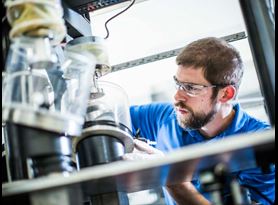
Element has the necessary facilities to perform large-scale cadaveric biomechanical testing. With a dedicated surgical suite, large-capacity freezers, and various types of mechanical testing frames, we can handle multiple test setups at once. Element’s prep room allows for specimens to be prepared in an independent temperature and humidity-controlled environment, and the testing laboratory is equipped with surgical instruments, surgical personal protective equipment (PPE), fume hood, dissection tables, and various other items. The surgical suite allows surgeons and researchers to come on-site and prepare the cadaveric specimens, perform the testing, and evaluate the results.
While having commercially available test frames such as MTS, Instron, and Bose is very useful and preferred for most testing, this is not always the case for cadaveric biomechanical testing. Cadaveric tests can be highly customized and involve complex setups. Our Engaged Experts have experience in building custom test frames and adding testing axes to commercially available frames to apply various loads, including loads along different axes and multiple loading points in the same axis.
Kinematic motion capture and data collection
Understanding how a device can alter the human body is often a missed concept in the biomechanical testing community. Once a device has been implanted, the injured area may not perform in the same manner as before the surgery, or there could be migration of the implant over time. For example, after a patient receives a total knee or hip arthroplasty, their gait may be altered and cause damage to surrounding joints or the device.
To truly understand how a device alters the articulation of a joint or migration of a system, a motion capture system can be utilized to quantify the kinematics. The motion capture system allows for the tracking of various points over the entire construct. Element performs motion capture with our four-camera Optitrak System, capable of tracking precise data collected from various markers placed throughout the bone, limb, or joint, in six degrees of freedom while collecting data at 120 Hz. When combined with force and displacement measurements, the function can be evaluated and quantified.
Conclusion
The objective of biomechanical testing is to determine how an orthopedic device will behave when implanted into a cadaveric specimen, closely simulating actual function in the human body. The testing allows for an overall look at how the device and body will behave when implanted and highlights potential resulting failure modes. Understanding these failure modes allows surgeons to determine indications for use, appropriate redesigns, and exclusion criteria for the device. While medical device companies work on making devices lighter, stronger, and safer, the resulting stresses created on the body can often be overlooked. Element offers a highly skilled team with a background in biomechanical testing of cadaveric specimens and the necessary resources to perform the testing in a safe and timely manner.
For more information about our biomechanical testing or other medical device testing services, contact our Engaged Experts.
Find related Resources
Related Services

Test Protocol for Medical Devices
A protocol and plan will mitigate your risk, prevent confusion, set clear expectations, and preserve the necessary information for future reference and use.

Preventing Fretting Corrosion of Orthopedic Devices
The human body is a corrosive environment that can compromise the performance of orthopedic devices. The test methods outlined in this article aid in preventing fretting corrosion in your device designs.
Medical Device Testing
As a comprehensive testing partner, you’ll enjoy the benefit of a single supplier source for all of your testing needs, from mechanical testing and environmental simulation to EMC and wireless device testing.
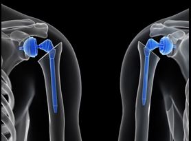
Preventing Glenoid Component Failure in Total Shoulder Replacement
Before total shoulder replacement devices are brought to market, they must be subject to various rounds of testing to prove that they are safe for use.